Thursday, December 3, 2009

Final Report: Overuse Injuries - A growing trend?

Biotechnology in Sports Medicine is becoming increasing relevant to young athletes. Throughout our blog, we have explored preventative techniques and treatment options for three main injuries associated with baseball, basketball, and rowing. We have noticed a growing trend in early specialization in specific sports, and there seems to be mounting pressure on today’s youth to perform at a higher level compared to previous generations. Additionally, because of the competitive nature of college admissions, people now see sports as a ticket to highly selective universities. This added focus on one sport has increased the prevalence of sports-related injuries, three of which we focused our research on: rotator cuff tears, ACL injury, and lumbar disc herniations.

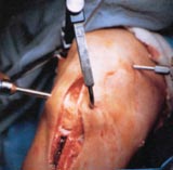
Rotator cuff tears sustained playing baseball are most often caused by overuse due to the repetitive throwing motion. These are complicated injuries because not only do they sideline athletes, but they strongly impair daily life. A friend, a former varsity athlete, just sustained a full thickness rotator cuff tear and is in extensive physical therapy to even be able to hold her child in the future. Rotator cuffs are most often repaired surgically and technology has improved recovery rates and surgical outcomes. The Opus Knot Fixation System is an example of a new biotechnology used in the surgical treatment of shoulder injuries, and may become more widely available in the future. On the flip side, because of the effectiveness of surgery, younger and younger patients are opting for surgery instead of more conservative treatments. Our blog also investigated the conservative treatment alternatives for these injuries. Conservative approaches include activity modifications, and oral non-steroidal anti-inflammatory medications – these treatments have been proven effective in the treatment of shoulder injuries.

Many elite Basketball players have sustained or will sustain an ACL injury. ACL injuries have become so prevalent that The American Academy of Orthopaedic Surgeons and the National Athletic Trainers' Association has taken a proactive stance through public service announcements to make athletes aware of the importance of preventing these injuries. ACL tears are more commonly found in girls both because of their gender, and because they are genetically pre-disposed (the COL5A1 gene) to the injury. There are a variety of techniques being developed both to improve and change surgical techniques. An example of this is the use of bone marrow stimulation to regenerate growth of the torn ACL – one day this technique may be able to replace ACL reconstruction.

Overuse injuries in rowing most commonly manifest themselves in the lumbar region. The rowing stroke includes a significant amount of lumbar flexion, which increases with fatigue. Over time this leads to back injuries, including lumbar disc herniation. Our group was particularly interested in these injuries, as two of our bloggers are NCAA rowers, and have experienced overuse injuries. On the extreme end, one rower had a transforaminal epidural steroid injection, and a medial nerve block to treat a lumbar disc herniation. As an example of a more conservative treatment another rower underwent bone stimulation, a new technology that has been used to treat stress fractures (commonly found in the ribs of rowers). With respect to conservative treatments and prevention – the volume of training required for rowing makes prevention of overuse injuries difficult. Usually prevention focuses on improving core strength and flexibility, which may decrease the probability of an injury occurring.

Exercise is an integral component of personal health. Given these issues with overuse, it is becoming necessary to find alternative forms of training. An athlete’s training schedule should include exercise which allows the body to rest from the repetitive nature of certain sports, and which increases the strength of a diverse group of muscles (some of which they would not exercise during regular training.)

Overall our blog sought to focus on the development of biotechnologies and prevention methods to alleviate sports-related injuries. We hope that both coaches and athletes will continue to investigate new solutions, such as those discussed in our blog, to improve the overall health of athletes, as well as to decrease the incidence of sports-related injuries.

Monday, November 23, 2009

A Less Invasive Treatment for Disc Related Lower Back Pain? (SpineCATH IDET)


Chronic back pain is commonly caused by fissures in the intevertebral discs of the spine. This injury has been followed by this blog, as it is commonly found in rowers, due to the stress they experience on their lower backs. Surgery to correct this problem comes with risks, long recovery periods, and other hurdles, and is not the only available option. The articles linked to below examine a non-surgical technology developed called Intradiscal Electrothermal Therapy (SpineCATH IDET). The article linked here comes from Science Daily, and discusses the technology, which started being widely available to patients around 2001.

http://www.sciencedaily.com/releases/2001/01/010124074916.htm

How does this procedure work? It is a simple treatment done under only a local anesthesia or mild sedation. A needle guided by x-ray is inserted into the afflicted disc, and then a catheter is passed through it and into position. The catheter is then heated, applying heat to the disc. This triggers a significant reaction in the disc causing the disc wall to heat up, and the fissures to contract and close. Along with this, the nerve endings may be desensitized, and/or the bulge of disk material can be reduced.

Chronic lower back pain is not exclusively found in athletes, in fact Science Daily reports in the article that one in five Americans suffer from back pain, and that non-surgical treatments are actually the most common. Technology like this is important because it offers athletes, and countless other people who experience disc-related back pain, a treatment option that is minimally invasive, and that also differs from more traditional non-surgical methods such as physical therapy and pain medications. It is great to see technology being applied not only to surgical procedures, but also to methods that are less invasive, and thus often carry fewer risks.

While there is still a recovery time required for the disk to heal (stated at 12-16 weeks), in the article below Dr. Amundson explains how a typical patient might be sent home after the procedure “with a Band-Aid over the needle insertion site.” That sounds a whole lot better than a large surgical incision.

For a discussion of the SpineCATH IDET procedure, possible side effects, and criteria for treatment look at the following article for a question and answer session with Dr. Glenn M. Amundson, an Orthopedic Surgeon about IDET:

http://www.spineuniverse.com/displayarticle.php/article217.html

Sunday, November 22, 2009

Advances in Biotechnology


http://rochesterchiro.files.wordpress.com/2008/09/drx9000.jpg

Advances in Biotechnology Improve Clinical Outcomes for Spinal Decompression


Many people suffer from back pains. Rowers in particular suffer from lumbar disc herniations, an uncomfortable spinal condition. A common solution is spinal decompression, but the procedure has many drawbacks. The purpose of the study by Gionis et al. was to determine the efficacy of recent advances in biotechnology used for spinal decompression. The DRX9000, a novel piece of equiptment recently approved by the FDA, was used in this study. Patients received twenty treatments of spinal decompression over a six-week course of therapy. Results showed that treatment was successful in 86% of the 219 subjects. These patients reported near complete resolution of pain, lumbar range of motion, and recovery of any sensory or motor loss.

Progress is the field of biotechnology is constantly under-valued; many people don't understand the science and judge based off of limited information. I feel it is important to pursue biotechnological solutions to common problems. If we don't we would miss out on such devices as the DRX9000 which led to very positive results as a treatment for disc herniation. Biotechnological advances in this area could further improve the results of treatments.

A Study on Strategies for Noncontained Lumbar Disc Herniations

The rowing stroke places a lot of stress on the lower back, thus leaving many competitive rowers dealing with lower back injuries. Although many young rowers present with bulging discs, annular tears, and inflamed joints, these problems often do not become surgical until later in life. A recent article in The Journal of Korean Neurosurgical Society discusses many implications of biotechnology in revolutionizing lumbar disc herniation surgery. Entitled “Strategies for Noncontained Lumbar Disc Herniation by Endoscopic Approach: Transforaminal Suprapedicular Approach, Semi-Rigid Flexible Curved Probe, and 3-Dimensional Reconstruction CT with Discogram”, Dr. Chae et al discuss the combination of imaging and surgical technology to increase the success rate and the ease of this particular surgery.

When a patient is diagnosed with a noncontained disc hernation, there is a distinct probability that the inside of the disc has fragmented and possibly moved around in the spine. With these types of herniations, the typical surgical methods are not always effective because of the placement of the disc fragments. Dr. Chae et al explain that they used 3D reconstruction CT images to determine the severity of the disc herniation and whether or not the fragments were contained. This allowed them to see whether or not the use of a semi-rigid flexible curved probe, made by RZ Medizin Technik GmBH in Germany, would help to extract the disc material so that the endoscope could reach the material and remove it from the body. The use of this type of probe allows the surgery to remain endoscopic (an incision of only 1 cm) even when the disc fragment has migrated into a place where it cannot be reached by the endoscope alone. In this study, 94.77% of the 145 patients had satisfactory results, and within that group, 99 patients reported excellent results.

Because of the variation in types of disc herniations, it is often difficult for doctors to determine what type of approach will be necessary to remove the disc fragment. This can make surgeries more difficult and possibly increase recovery time. Yet, according to this study, the use of 3D reconstruction CT allows surgeons to see exactly where the disc herniation has occurred and can determine what type of surgical technique and instrumentation will be necessary. Surgical tools like the semi-rigid flexibly curved probe can help mediate problems like disc fragment migration and also keep the surgery as simple as possible for the patient. These types of innovations can help to decrease the likelihood of needing revision surgery in the future by making sure that all of the disc fragments were successfully removed the first time around. Most athletes, especially rowers who place a lot of strain on their lower back, and commonly suffer from herniated discs, would welcome innovative techniques like this.

http://www.jkns.or.kr/htm/fulltext.asp?no=0042009141

Rowing Injuries: Possible Effects of Lumbar Flexion and Back Muscle Activity


Source

Low back pain is one of the most common injuries seen in rowers. Within the rowing stroke, although the legs are the main component of power, the back extensor muscles are pivotal in the regulation of flexion and extension of the lumbar spine. It has been suggested that the range of lumbar flexion over the course of the stroke may contribute to injury. In accordance with this, prior studies have shown that bending increases as people come near the top of their range in lumbar flexion, this consequently increases stress on spinal structures.

The study, “The Effects of Repetitive Motion on Lumbar Flexion and Erector Spinae Muscle Activity in Rowers” investigated the changes in lumbar flexion and back muscle activity throughout the course of a rowing trial on an ergometer. It was published in Clinical Biomechanics in May of 2003 and is available online. The study involved 16 young adult rowers with an equal sex ratio, of whom none had any medical history of lower back pain.

The study measured lumbar flexion and muscle activity at two parts in the rowing stroke and at three separate times during the trial (at 20%, 60%, and 95%). Motion analysis was used to record lumbar flexion and was then compared to each individual’s initial total range of lumbar motion. EMG recordings were used to record muscle activity in the multifidus, ilicostalis lumborum, and longissimus thoracis; they were then compared to each individual’s initial maximum voluntary isometric contraction readings.

The study concluded that rowers have an initial high level of lumbar flexion that continues to increase over the duration of a rowing trial. The changes in EMG recordings provided indirect evidence of fatigue in the back extensor muscles, which may also contribute to the increase in lumbar flexion. They found no significant difference between genders.

Although the study has flaws in it’s design, the article was very effective in showing the importance of conducting further research in the area and how beneficial it could be to injury prevention in rowing. Study weaknesses included the small sample size as well as the possibility of crosstalk in EMG recordings. As a rower, I found this article to be very interesting. A few things I thought worth noting, which the article failed to discuss when it compared the ergometer to actual rowing, were the possible difference in the fluidity of the stroke when moving on water and the perceived weight of the handle during the drive.

See the Article Here!

Olympic Rower Undergoes Micro-Discectomy


In September 2008 Australian triple Olympic gold medalist rower Drew Ginn underwent micro-discectomy spinal surgery to repair a lower back problem caused by rowing. After winning the gold medal in the summer 2008 Beijing Olympics, Ginn needed to get surgery. He almost had to drop out of the Beijing Olympics due to a ruptured disc. He was experiencing nerve pain and ultimately discovered he had ruptured his disc, and that a fragment had broken off and been lodged in the cavity behind the disc. He pushed through the 2008 Olympics with the pain, but once it was over had a micro-discectomy to repair the ruptured disc in the L4/L5 region of his lower back. The pain had become so overbearing that surgery was Ginn’s only option. The pain in his lower back was moving down to the sciatic nerve in his leg. Micro-discectomoy’s are performed for herniated lumbar discs and effectively treat leg pain or radiculopathy. The recovery time for this procedure is about 6 weeks; Ginn had to stay in bed, only walking when movement was necessary. This is a significant amount of a time for an athlete in particular to have to remain inactive.


There are some risks associated with micro-discectomy’s such as dural tear, nerve root damage, infection, and bladder incontinence. Ginn experienced some nerve damage and deactivation of muscles around his hip, back, and right leg. The damage could be permanent but it takes about 6 months to determine that. Complications for micro-discectomy's are rare.


Ginn has had a long history of back injuries during his rowing career. He missed the Sydney Olympics due to a back injury and went through a rehabilitation process in 2000. This article provides a great case study of the dangers and severity of injuries associated with rowing athletes.

Monday, November 16, 2009

Check out this video of a Basketball player tearing their ACL

http://www.youtube.com/watch?v=84OUUz3u7Lc&feature=player_embedded#at=16

Collagen-Platelet Composites Found to be Effective in ACL Repair


In 2006, an article in Science Daily reported on a promising technique that could one day replace ACL surgery. The technique described in the article involved injecting collagen gel, enriched with blood platelets from the patient, into the area where the ACL tear had occurred. This collagen would then it was theorized, act as a bridging material for the torn ligaments to fill. Meanwhile, the platelets would supply growth factors that would support this bridging. This technique had only been explored in animal models at the time, but nonetheless was exciting. Dr. Martha Murray, who led the team conducting the study, hoped that the technique would provide a less-invasive, more effective form of ACL repair, and that the collagen gel could eventually be used to regenerate cells damaged by osteoarthritis.

Building on this information, this past March 2009 researchers published an article in the American Journal of Sports Medicine that shows further promise for the utility of collagen-platelet composites. The study, titled “Collagen-Platelet Composites Improve the Biomechanical Properties of Healing Anterior Cruciate Ligament Grafts in a Porcine Model,” compared surgical outcomes in porcine subjects. The pigs were either given a traditional ACL repair, or an ACL repair with a collagen-platelet composite (CPC) around the graft site. Results are very promising. The CPC was well tolerated, as it was pre-treated with a pepsin product to help reduce rejection. Animals who received this treatment showed a stronger recovery from their ACL repair. Those who received the CPC had no observed necrosis in the graft, and had a lower level of knee laxity (instability) post-op.

The results of this study are exciting, and reinforce the thought that collagen support in ACL repair could hold promise as a future treatment in human patients. There are however a few caveats, since the grafts were used in animals they were not sterilized prior to grafting. There is no way of knowing whether the sterilization process that would be necessary to put an allograft into a human would reduce the efficacy of the treatment. Additionally, there remains the chance that the process of wound recovery, and the physiological differences between the porcine model and that of a human, contain variation that could influence the extent to which the treatment is effective. Moreover, the technique has not yet been tested in a human.

The results of the study still show that although the enhanced surgical graft was effective, that there is no substitute for a healthy, injury-free ACL. Thus, even with such exciting technologies beginning to emerge, prevention should still remain a focus as medicine moves forward.


The articles discussed in this post can be found at:

Sunday, November 15, 2009

Study Shows a Potential Future Alternative to ACL Reconstruction

Source

One of the most commonly treated knee injuries is an anterior cruciate ligament tear. It has been documented in the past that the ACL has a very limited ability to heal itself; this has made reconstructive surgery the standard treatment for ACL tears. In reconstructive surgery, the torn ACL is removed and replaced with a tissue graft often from the hamstring or patellar tendon. Another surgical option is primary repair, in which, the injured ACL is sutured back onto the residual ACL. This technique is not commonly used because it has been shown to result in continual laxity and instability of the knee.

The study, “Primary Repair Combined With Bone Marrow Stimulation in Acute Anterior Cruciate Ligament Lesions” hypothesized that primary ACL repair with additional bone marrow stimulation could successfully restore stability and function in the knees of athletes with partial tears. It was published in March of 2009 in the American Journal of Sports Medicine and can be found online on their website. The study consisted of 26 athletes, with a mean age of 26.6 years, all of whom were able to undergo surgery within 4 weeks. All participants in the study had athroscopically confirmed partial ACL tears and underwent a primary repair surgery in which there was also bone marrow stimulation of the ACL femoral attachment area. They also had to undergo a specific rehabilitation program post-op. After surgery and rehabilitation, only 19% of the patients did not return to the same initial level of sports activity, in more than half of these patients the determining reason was personal and not due to physical impairment.

The study’s use of bone marrow stimulation builds upon the notion that the use of stem cells in medicine may be at large in the near future. The stimulated cells were mesenchymal stem cells, which have healing capabilities because of their ability to differentiate into specific tissue.

The study concluded that primary repair of an acute ACL tear paired with bone marrow stimulation can successfully restore the knee’s function and stability. Unfortunately the small group size, strict experimental criteria, and lack of a control group/randomization make the study statistically insignificant. Instead, by revealing the possible use of natural healing in ACL injuries, the study serves as a window into what may eventually replace ACL reconstruction in the future.

Although this study has many flaws, it effectively promotes the benefit that may come out of further research into alternative ACL surgery. If successful, this surgery would be a great alternative to total ACL reconstruction. It would theoretically eliminate the decreased proprioception that ACL-reconstructed knees often suffer from as well as harvest site morbidity and harvested tendon weakness.

See the Online Article Here!

Patellar Tendon Versus Hamstring Tendon Autografts for Reconstructing the ACL

ACL (anterior cruciate ligament) injuries are very common among athletes and non-athletes alike, but sports like basketball tend to have a very high incidence rate of ACL tears. Jumping and twisting both put unnatural strain on the knee ligaments, and if a basketball player lands incorrectly, he/she can be out for the season and will be faced with a series of complicated decisions. Some ACL tears do not require surgery, but in order to return to athletics, it is usually necessary to surgically reconstruct the ACL. ACL reconstruction surgery is done using one of three types of grafts to replace the torn ligament: a piece of the patient’s patellar tendon, a piece of the patient’s hamstring tendon, or an allograft (usually a cadaveric Achilles tendon). Autografts have become the go-to surgical option for numerous reasons, but there has been a longstanding discrepancy between the use of patellar and hamstring autografts.

In August of 2009, The American Journal of Sports Medicine published a meta-analysis on individual patient data entitled “Patellar Tendon Versus Hamstring Tendon Autografts for Reconstructing the Anterior Cruciate Ligament”. This study looked at data from 6 previously published clinical trials and used two different knee stability tests to analyze the effectiveness of both grafts. Dr. Blau et al used the positive pivot-shift test to characterize knee instability and the positive Lachman test for instability on a post-ACL reconstruction knee. The bone-patellar-tendon bone autograft had significantly fewer positive pivot-shift tests, even when adjusted for age and gender. Because there are so many factors contributing to how individual knees recover and perform, some surgical options work better for some, but this study determined that, despite differences, pattelar tendon autografts are superior to hamstring grafts.

Orthopedic surgeons advise their patients based on a variety of factors, but as research on graft superiority becomes more definitive, the reconstruction procedure should become more standardized. For athletes like basketball players in which these injuries are so common and for whom stability plays an important role in their sport, bone-patellar-bone tendon autografts may be the answer for ACL reconstruction.

http://ajs.sagepub.com/content/early/2009/08/18/0363546509333006.long